Researchers create a way to visually sort blood cell data
For more than five decades, researchers wanting to learn more about cellular composition and cellular physiology used a process called flow cytometry to examine the physical and chemical aspects of blood and other kinds of fluid. It involves passing the substance through at least one laser.
Any fluorescent molecules that are present emit fluorescence, while other particles show light-scattering properties.
A computer then processes those associated light signals and allows finding out about components related to a large number of cells in a relatively short span of time.
However, an international team comprised of researchers from numerous universities came up with a more advanced method they called Image-Activated Cell Sorting (IACS).
Parts of the process are the same as flow cytometry, but most importantly, IACS creates two-dimensional images from cell data using deep learning.
How Does It Work?
The scientists who worked on the project report that IACS allows for sorting cells based on their global phenotypic profiles as well as the morphological properties and where the cells are in space. While other hematology techniques might place more emphasis on removing the need for a diluent or liquid reagent, IACS classifies the cells by spatial and morphological properties not possible through traditional flow cytometry.
The IACS system took six years to create between the design of the platform and its subsystems, plus a two-year testing process involving blood cells and algae.
The cell-sorting process starts when researchers choose desired criteria and insert a tube filled with suspended cells into the IACS injection point.
Then, each cell gets imaged individually under a microscope. It’s at this stage when the intelligent decision-making abilities of the IACS process come into play. The system analyzes the collected data made up of the information from the individualized cells.
It then determines which cells -- if any -- match the initially chosen criteria. Those that do match get separated from the rest of the cells.
As such, a completed IACS process features two tubes of cells. One contains sorted cells, and the other has unsorted cells. The last step is evaluating the contents of each cell with an optical microscope to check for quality.
This big data-driven approach permits scientists to evaluate substantial quantities of cells much faster than previous methods allowed.
Moreover, the degree of accuracy is comparatively higher, which could potentially lead to discoveries that wouldn’t be possible without this technology.
A Quick and Multifunctional System
A research paper about the scientists’ findings says that IACS acquires images, processes them and makes decisions based on the data all within 32 milliseconds. Additionally, users can search and sort through image-based cell classifications at a rate of approximately 100 cells per second.
Doctors commonly prescribe complete blood counts (CBCs) for patients to diagnose conditions, monitor the effectiveness of ongoing treatments and gauge overall health, among other things. Advancements in hematology capabilities allowed physicians to obtain results while patients waited on site. Other technologies permit performing CBCs during fieldwork or in different demanding environments.
The dozens of scientists who made IACS a reality say their technology could be yet another groundbreaking improvement in hematology as well as other fields. For now, the IACS system looks only at individual cells. However, scientists believe they could adjust the platform and make it work with larger items, such as tissue fragments and even entire organisms.
A Highly Scalable Solution
Some emerging technologies aren’t as versatile as first imagined because they take a prohibitively long time to adjust for new uses. However, the researchers familiar with the IACS discussed its adaptability at length in a recognized scholarly publication. For example, they clarified how it would be possible to use the IACS for different purposes by changing the imaging technology used or applying computational additions like GPUs or CPUs.
They also mention that because this is a machine-learning-powered technology, it’ll get smarter with use. Moreover, users could take the sorted images of cells and use that content to improve future outcomes. For example, since this method depends on big data for both accuracy and speed, it could draw attention to rare kinds of cells that scientists wouldn’t spot through older cell analysis methods.
Once the IACS system gets exposed to those cells that aren’t often seen, scientists could teach algorithms with the cells the system found. The associated things experts learn might spur the development of new medicines or help researchers identify and target populations with unusual cell characteristics.
Offering an Open-Access Approach to Further Study
The IACS system is in Tokyo, Japan, and it’s so complex that people cannot easily recreate the system elsewhere. However, the original team wanted to make the technology available to other researchers and created a survey anyone can access and fill out for a chance to collaborate with the IACS researchers for further work. Plus, a Japanese startup called Cybo aims to commercialize IACS and appears to be focusing primarily on the technology detailed above.
Big Data and Related Technologies Open up New Opportunities
The discussion of IACS and how it uses deep learning to speedily and accurately categorize cells via image-based means offers substantial potential for biologists and other professionals working with cells.
This advancement already shows promise, but the full potential of the progress will likely be realized only later.
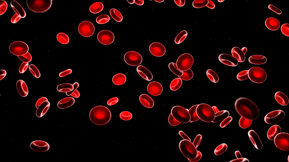
Photo Credit: Sebastian Kaulitzki/Shutterstock
 Kayla Matthews is a senior writer at MakeUseOf and a freelance writer for Digital Trends. To read more from Kayla, visit her website productivitybytes.com.